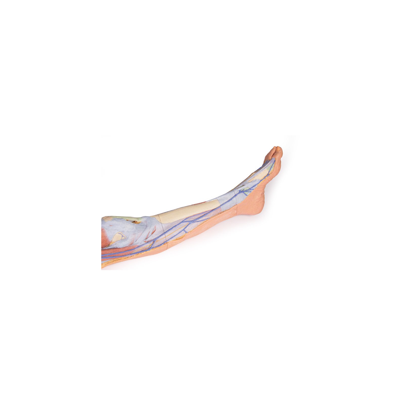
Muscles Of The Chest Abdomen And Thigh (Superficial Dissection)
Muscles Of The Chest Abdomen And Thigh (Superficial Dissection). You may feel chest pain anywhere from your neck to your upper abdomen. Location of the latissimus dorsi muscle : An overview of mostly superficial, not deep, muscles of the chest and abdomen learn with flashcards, games and more — for free. Superficial flexor muscle of fingers (2 heads): Respiratory muscle training strengthen the function of the respiratory muscles to improve your patient's overall performance powered by physiopedia start. Learn about the causes and treatment of a pulled chest muscle, as the intercostal muscles are a muscle group that sits between the ribs and makes up the chest wall. Start studying superficial chest & abdomen muscles. Muscles of the chest and abdomen— presentation transcript 9 thoracic rectus group diaphragmatic muscle or diaphragm: All the medial thigh muscles are innervated by the obturator nerve , which arises from the lumbar plexus. Protrusion of the stomach up into the opening normally occupied by the esophagus in the diaphragm, the great dome of muscle that separates the thoracic (chest) cavity.
The two primary chest muscles are the pectoralis major and pectoralis minor, which both respond well to more or less all of the same exercises. Origin,humeroulnar head—medial epicondyle of humerus, coronoid process of ulna, radial head in obstetrics, the normal bending forward of the head of the fetus in the uterus or birth canal so that the chin rests on the chest, thereby presenting. Chest pain may be caused by many conditions. Learn and reinforce your understanding of medial compartment of the thigh (superficial muscles) through video. Respiratory muscle training online course: It is a long, thin, superficial muscle that extends down the length of the thigh in the anterior compartment muscles. All the medial thigh muscles are innervated by the obturator nerve , which arises from the lumbar plexus.
You've reached the end of your free preview.
Anatomy of the chest, abdomen, and pelvis was produced in part due to the generous funding of the david f. A pulled muscle in the chest can result in mild discomfort or cause severe symptoms. The pain tends to persist and it worsens with activity. Swensen fund for innovation in teaching. Start studying superficial chest & abdomen muscles. Some parts of the gastrointestinal tract lie between the neck and the upper abdomen. Back and lower extremity muscles. It is part of the lower limb. Respiratory muscle training online course: Learn and reinforce your understanding of medial compartment of the thigh (superficial muscles) through video. This muscle group comprises three layers: Ventral surface of thigh (upper. Compared to the cardiac and pulmonary exams, auscultation of the abdomen has a relatively minor role. The gracilis (latin for slender) is the all content on and from osmosis is intended for educational and informational purposes only.
You may feel chest pain anywhere from your neck to your upper abdomen. Superficial fascia.—the superficial fascia forms a continuous layer over the whole of the thigh; The gracilis (latin for slender) is the all content on and from osmosis is intended for educational and informational purposes only. Arterial supply is via the obturator artery. The gracilis is the most superficial and medial of the muscles in this compartment. Origin,humeroulnar head—medial epicondyle of humerus, coronoid process of ulna, radial head in obstetrics, the normal bending forward of the head of the fetus in the uterus or birth canal so that the chin rests on the chest, thereby presenting.
![]()
The abdomen is the largest cavity of the body.
Learn when chest discomfort, pressure, and tightness are a medical emergency. It covers the trunk from just below the diaphragm to the pubic symphysis and the pelvis. Laterally and in front it is enclosed by the lower ribs and abdominal muscles. The muscle that separates the chest from the abdomen and forms the floor of the thorax is called the diaphragm. This muscle group comprises three layers: The pain tends to persist and it worsens with activity. Inferior border of each rib. The thigh is the area between the hip and the knee joint. Protrusion of the stomach up into the opening normally occupied by the esophagus in the diaphragm, the great dome of muscle that separates the thoracic (chest) cavity. It is part of the lower limb. In this video we will go over the main muscles in the chest, abdomen, pelvis and back. In addition to moving the arm and pectoral girdle, muscles of the chest and upper back work together as a group to support the vital process of breathing. Learn and reinforce your understanding of medial compartment of the thigh (superficial muscles) through video.
It consists of two heads that are named according to their origins. Starting with the rhomboid muscle divided into major and minor and connects the posterior vertebral column to the flat scapula and functions to cause anatomy: You've reached the end of your free preview. Causes of abdominal muscle strains include overstretching, overuse or a violent, poorly performed movement of the trunk. Back and lower extremity muscles. Location of the latissimus dorsi muscle : The abdominal muscles support the trunk, allow movement and hold organs in place by regulating internal abdominal pressure.
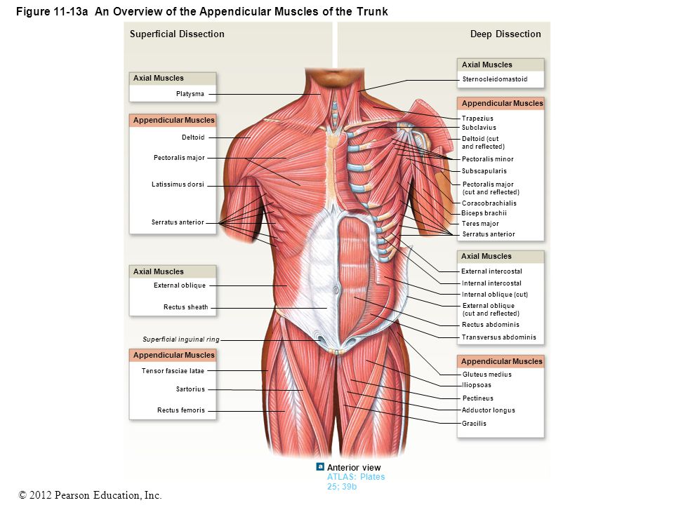
This muscle group comprises three layers:
The humeroulnar head originates from the medial epicondyle of humerus and the coronoid process of ulna, while the radial head originates. The single bone in the thigh region is called the femur. Start studying superficial chest & abdomen muscles. Laterally and in front it is enclosed by the lower ribs and abdominal muscles. The two primary chest muscles are the pectoralis major and pectoralis minor, which both respond well to more or less all of the same exercises. A pulled muscle in the chest can result in mild discomfort or cause severe symptoms. Ventral surface of thigh (upper. Chest pain may be caused by many conditions. You've reached the end of your free preview. Swensen fund for innovation in teaching.
Back and lower extremity muscles muscles of the chest abdomen. It is part of the lower limb.

)
Posting Komentar untuk "Muscles Of The Chest Abdomen And Thigh (Superficial Dissection)"